
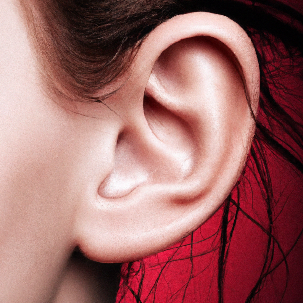
The ear is an incredible sensory organ that plays a vital role in our ability to hear and maintain balance. Divided into three parts – the outer ear, middle ear, and inner ear – each section contributes to the complex process of sound perception. Starting with the outer ear, which includes the auricle and external ear canal, sound waves are funneled towards the middle ear through the help of keratinized stratified squamous epithelium. The middle ear, connected to the nasopharynx by the Eustachian tube, contains three small bones known as ossicles that transmit vibrations from the tympanic membrane to the inner ear. Here, in the inner ear’s cochlea and vestibule, fluid waves are converted into nerve signals responsible for our ability to hear and maintain balance. The ear is a intricate and impressive organ that relies on a series of complex processes to achieve its functions.

Overview of the Ear
Introduction
The ear is a complex sensory organ responsible for hearing and balance. It plays a crucial role in our daily lives, allowing us to communicate, enjoy music, and stay aware of our surroundings. In addition to its sensory functions, the ear also plays a role in regulating our equilibrium, helping us maintain balance and coordination.
Structure of the Ear
The ear is divided into three main parts: the outer ear, middle ear, and inner ear. Each part has its own unique structures and functions that contribute to the overall functioning of the ear.
Functions of the Ear
The primary function of the ear is to detect and transmit sound waves to the brain, allowing us to hear and understand sounds. Additionally, the ear is responsible for maintaining our sense of balance and equilibrium, ensuring that we can move and navigate our environment without falling or experiencing dizziness.
Anatomy of the Ear
Outer Ear
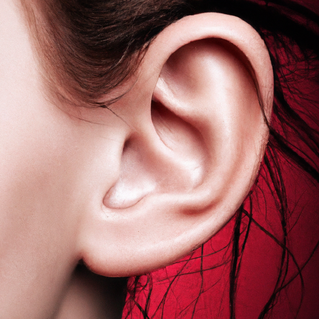
The outer ear is the visible part of the ear that we commonly refer to as the earlobe or pinna. Its main function is to collect sound waves and direct them into the ear canal. The outer ear consists of two main structures:
Auricle
The auricle, also known as the pinna, is the outermost part of the ear that is visible on the side of the head. It is made up of cartilage covered by skin and helps to direct sound waves into the ear canal.
External Ear Canal
The external ear canal, also known as the auditory canal, is a tubular structure that connects the auricle to the middle ear. It is lined with keratinized stratified squamous epithelium, which helps protect the ear from foreign particles and keep it clean.
Epithelium of the Outer Ear
The epithelium of the outer ear plays a crucial role in maintaining the health and function of the ear. It protects the underlying tissues from infection and damage, while also producing cerumen, commonly known as earwax, which helps to lubricate and protect the ear canal.

Middle Ear
Connection to Nasopharynx
The middle ear is connected to the nasopharynx by a small tube called the Eustachian tube. This tube helps equalize the pressure between the middle ear and the external environment, preventing discomfort and damage to the eardrum.
Ossicles
The middle ear contains three small bones, known as ossicles, which are the smallest bones in the human body. The ossicles include the malleus, incus, and stapes. These bones work together to amplify sound vibrations and transmit them from the tympanic membrane (eardrum) to the inner ear.
Transmission of Vibrations
The ossicles play a crucial role in transmitting sound vibrations from the eardrum to the inner ear. When sound waves reach the eardrum, they cause it to vibrate. These vibrations are then transmitted through the ossicles to the oval window, a membrane-covered opening that leads to the inner ear.
Inner Ear
Cochlea
The cochlea is a spiral-shaped, snail-like structure located in the inner ear. It is responsible for converting mechanical vibrations into electrical signals that can be interpreted by the brain as sound. The cochlea contains specialized hair cells that detect these vibrations and transmit them to the auditory nerve.
Vestibule
The vestibule is another important structure in the inner ear. It plays a crucial role in our sense of balance and spatial orientation. It contains small organs called otoliths, which detect changes in head position and movement, helping us maintain our equilibrium.
Conversion of Fluid Waves to Nerve Signals
In the inner ear, fluid waves generated by sound vibrations are converted into nerve signals that can be understood by the brain. This conversion process occurs in both the cochlea and the vestibule, where specialized cells translate mechanical energy into electrical signals that can be transmitted to the brain.
Functioning of the Ear
Transmission of Sound Waves
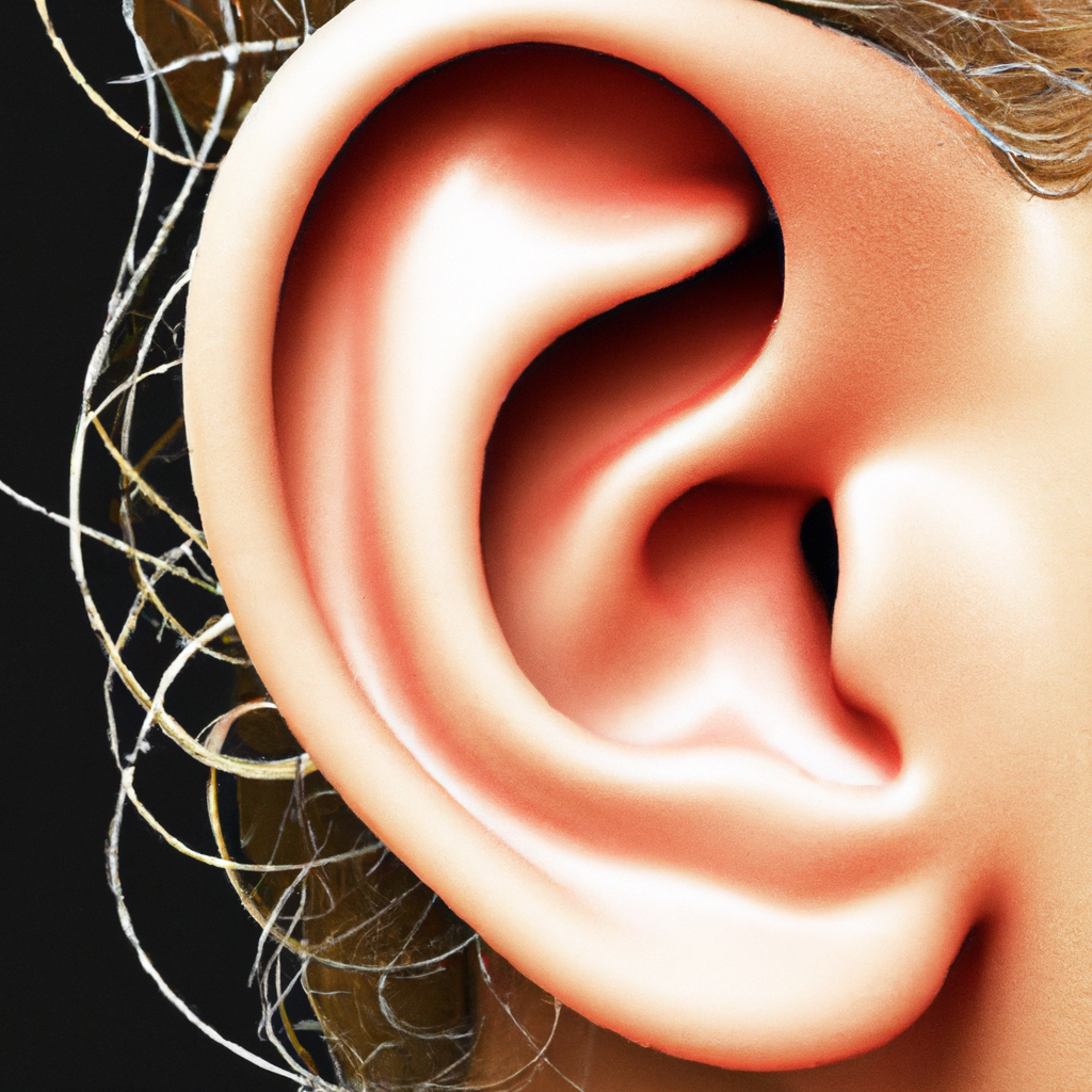
The functioning of the ear begins with the transmission of sound waves from the environment to the tympanic membrane. These sound waves are collected by the auricle and funneled into the external ear canal. From there, they travel to the eardrum, causing it to vibrate.
Modulation in the Middle Ear
The vibrations of the eardrum are then transmitted to the ossicles in the middle ear. The ossicles amplify and modulate these vibrations, allowing for finer control over the volume and frequency of the sound waves.
Conversion of Mechanical Energy to Electrical Energy
In the inner ear, the cochlea and vestibule convert the mechanical energy of the sound vibrations into electrical energy. This conversion is carried out by specialized hair cells that detect the vibrations and generate electrical signals.
Transmission of Information to the Brain
The electrical signals generated by the hair cells are transmitted to the auditory nerve, which carries them to the brain for interpretation. The brain processes these signals and allows us to perceive and understand sounds.
Histologic Observation of Ear Tissues
Preparation Techniques
To observe ear tissues at the microscopic level, various preparation techniques are used. These techniques include fixation, processing, embedding, and staining.
Fixation
Fixation involves preserving the ear tissues in a state as close to their living state as possible. This is usually done by immersing the tissues in a fixative, such as formalin, which stabilizes the cellular structures and prevents degradation.
Processing
After fixation, the tissues undergo processing, which involves dehydrating them and impregnating them with a substance that can be easily sectioned. This allows for thin slices of the tissues to be prepared for microscopic examination.
Embedding
Embedding is the process of embedding the dehydrated tissues in a solid medium, such as paraffin wax. This provides support and allows for thin sections to be cut for staining and examination under the microscope.
Staining
Staining is a crucial step in the histologic observation of ear tissues. It involves applying various dyes or stains to the tissue sections to enhance contrast and visibility. Common stains used in ear research include hematoxylin and eosin, trichrome, and immunohistochemistry.
Stains Used in Ear Research
Hematoxylin and Eosin
Hematoxylin and eosin staining is one of the most commonly used staining techniques in histology. Hematoxylin stains the nuclei of cells a dark blue color, while eosin stains the cytoplasm and extracellular matrix pink or red. This staining technique provides valuable information about the cellular structures and tissue organization in the ear.
Trichrome
The trichrome stain is used to highlight collagen and other connective tissues in the ear. It consists of three dyes: hematoxylin, which stains nuclei blue, acid fuchsin, which stains cytoplasm and muscle fibers red, and aniline blue, which stains collagen fibers blue. Trichrome staining helps visualize the distribution and organization of connective tissues within the ear.
Immunohistochemistry
Immunohistochemistry is a staining technique that allows for the detection of specific proteins or molecules within the ear tissues. Antibodies labeled with fluorescent or enzyme tags are used to bind to their target antigens, which can then be visualized under a microscope. Immunohistochemistry is particularly useful for studying the distribution and localization of specific molecules in the ear.
Microscopic Examination of the Ear
Structural Components
Microscopic examination of the ear allows for a detailed analysis of its structural components. This includes the different layers of the tympanic membrane, the three ossicles of the middle ear, and the intricate structures of the cochlea and vestibule in the inner ear.
Specialized Cells
Microscopy also allows for the visualization of specialized cells within the ear. These include hair cells, pillar cells, and neurons, which play important roles in the detection and transmission of sound and balance signals.
Hair Cells
Hair cells are specialized sensory cells found in the cochlea and vestibule. They have hair-like projections called stereocilia that detect sound vibrations and convert them into electrical signals. Microscopic examination helps to observe the arrangement and condition of these hair cells, providing valuable insights into hearing and balance disorders.
Pillar Cells
Pillar cells are found in the cochlea and provide structural support for the sensory hair cells. They help maintain the integrity and function of the cochlea and play a role in sound detection. Microscopic examination allows for the observation and study of pillar cell morphology and organization.
Neurons
Neurons are specialized cells that transmit electrical signals from the ear to the brain for processing. Microscopic examination can reveal the presence and organization of these neurons, providing insights into the neural pathways involved in hearing and balance.
Prevention of Hearing Loss
Public Health Measures
Hearing loss is a common problem that can have a significant impact on quality of life. Fortunately, many cases of hearing loss can be prevented through public health measures. These measures include raising awareness about the importance of hearing protection, implementing regulations to reduce noise exposure at workplaces, and ensuring access to affordable and high-quality hearing healthcare services.
Global Hearing Loss Statistics
Hearing loss is a global health issue, affecting people of all ages and backgrounds. According to the World Health Organization (WHO), approximately 1.1 billion people worldwide experience some degree of hearing loss. This represents about 15% of the global population. Furthermore, it is estimated that about half of all cases of hearing loss could be prevented through public health measures.
In conclusion, the ear is a remarkable sensory organ that allows us to hear and maintain our sense of balance. It is a complex structure composed of the outer ear, middle ear, and inner ear, each with its own unique functions and structures. Understanding the anatomy and functioning of the ear can help us appreciate the intricate mechanisms involved in hearing and balance. Through histologic observation and microscopic examination, researchers can gain valuable insights into the ear’s structure and specialized cells. By implementing public health measures and raising awareness, we can work together to prevent hearing loss and ensure that everyone can enjoy the gift of sound.
